This scan helps us to see the heart's arteries. We use it to look for any blockages or narrowing of the arteries. It also helps us to see any calcium deposits which may be a cause for concern.
This is a more specialist scan to assess the structure and function of the heart. It takes detailed pictures of the heart muscle, valves, and blood vessels. We are one of only three Trusts in the region to do this.
This is where we use a small tube to insert dye into your blood stream. We then take X-ray pictures of the heart to see how the dye moves around your heart. We do this in our cath labs on both hospital sites. If we find a problem, we can often fix it there and then.
An ECG shows us how your heart is working. It is an easy test to do. We put small sticky patches called electrodes on your chest, arms, and legs. These connect to a machine which shows us how fast your heart is beating and the rhythm.
We use this test to see how well blood flows to your heart muscle. We will measure your heart while you walk on a treadmill. Or we might give you a medicine to make your heart work harder. We use a dye and a special camera to see where the blood is flowing.
This is an ultrasound scan which shows us a moving picture of your heart. We can see the size and shape of your heart chambers and the thickness of your heart walls. It also shows how well the valves are opening and closing. We use this to keep an eye on various heart conditions and find new ones.
This is also known as a stress test. We look to see how well your heart works when you do physical activity. You will walk on a treadmill or ride an exercise bike while we monitor your ECG.
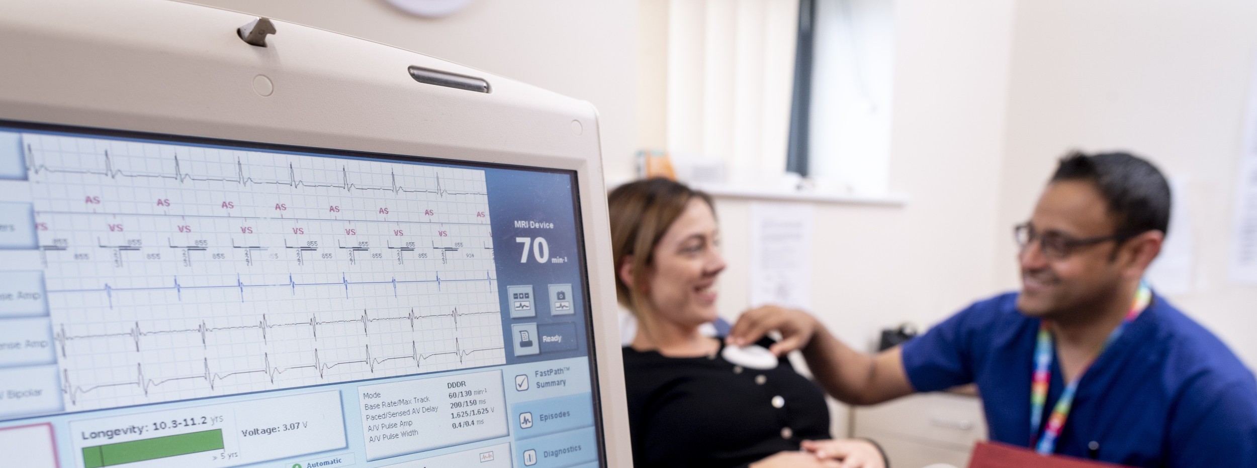
We may need to track how your heart works at home and in your normal life. We will ask you to wear a remote monitoring device to do this. We may also ask you to record and monitor your blood pressure.